Respiratory Sinus Arrhythmia: How Breathing Shapes Your Heart Rhythm
Yakiv Bilenko — editor · Updated October 5, 2026
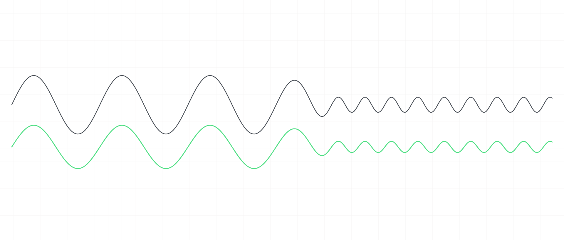
Respiratory sinus arrhythmia (RSA) is the normal rise in heart rate during each breath in and fall during each breath out. Despite the name, it is a feature of a healthy resting heart, not a rhythm disorder. It is carried largely by vagal regulation of the heart and is shaped by how slowly and deeply you breathe, by posture and by age. It does not by itself establish vagal tone, fitness or health.
Key points
- Respiratory sinus arrhythmia is the breath-by-breath rise and fall of heart rate: faster on the in-breath, slower on the out-breath.
- It is a normal feature of a healthy resting heart, not a heart rhythm disorder, despite the word arrhythmia.
- Vagal regulation of the heart carries most of it; how breathing and the baroreflex combine to generate it is still debated.
- It is larger with slow, deep breathing and when lying down, and smaller with fast breathing and during exercise.
- In short resting recordings it is the main source of heart rate variability, which is why breathing moves RMSSD so visibly.
- Because breathing itself changes its size, RSA is not a clean readout of vagal tone.
- Why the body has RSA is still a matter of hypotheses, and its size is not a diagnosis or a health score.
What is respiratory sinus arrhythmia?
Respiratory sinus arrhythmia (RSA) is the natural rise and fall of heart rate with each breath: the heart speeds up slightly as you breathe in and slows as you breathe out [S2] [S4]. "Sinus" means the beats still start in the heart's normal pacemaker; "respiratory" names the driver. The word "arrhythmia" here is a historical label for a varying rhythm, not a diagnosis — RSA is the rhythmic fluctuation of beat-to-beat intervals seen in healthy people at rest [S5].
Researchers recommend keeping the term for exactly this breath-linked speeding and slowing of the heart [S3]. It is the most visible part of heart rate variability — the beat-to-beat variation in the heart's rhythm — and this page is about that one component: what produces it, what changes its size and what it can and cannot tell you.
How does it work?
What is established. RSA is carried mainly by the vagus nerve: it is the respiration-driven speeding and slowing of the heart via vagal regulation [S1]. The mechanisms are understood to involve the brain's breathing drive and stretch reflexes from the lungs acting on the vagal outflow to the heart, and RSA becomes larger as that vagal influence on the heart grows [S7].
What is still a model or a debate. How exactly breathing and circulation combine to generate RSA is not settled. One long-running debate asks whether a central respiratory centre in the brainstem or the baroreflex — the blood-pressure feedback loop — predominantly generates it [S4]. One view holds that the coupling of blood pressure and heart intervals at breathing frequencies mostly reflects breathing acting on both, rather than baroreflex physiology [S5]; others give the baroreflex a larger role. The broadly accepted middle ground is that central, peripheral and mechanical elements interact to produce it [S4]. In practice, the page you are reading treats the vagal pathway as established and the relative weight of each generating mechanism as open.
Why slow breathing makes RSA so much larger — the link to resonance and the baroreflex — is a mechanism question of its own, planned for a separate page on breathing and HRV.
How is it measured?
RSA is read from the intervals between heartbeats recorded alongside breathing, or estimated from heart rate variability itself. In short resting recordings it is the primary source of the variation, especially with slow, paced breathing [S1] — which is why a breathing exercise visibly moves an HRV number. Among time-domain metrics, RMSSD typically gives a better assessment of RSA than related measures [S1]. The general rules for comparing such numbers — same device, same metric, comparable conditions, your own baseline rather than someone else's value — apply here as well.
What affects it?
- Breathing rate. RSA is small at fast breathing rates and large at slow ones [S5] [S4]. Typical resting breathing sits around 12–20 breaths per minute; as breathing slows, RSA grows, and it tends to peak around about 5.5–6 breaths per minute (around 0.1 Hz) — a value that varies from person to person [S4]. Finding your own rate is a practical question covered in find your resonance breathing rate.
- Breathing depth. A larger breath increases RSA, more so at slow rates [S4], and in a classic laboratory study its size rose in proportion to breathing volume [S6].
- Body position and state. RSA is largest during sleep, relaxation, slow deep breathing and when lying down, and smaller during exercise and anxiety [S4].
- Age. On average, HRV tends to fall with age — in our night-time RMSSD table the median drops by about 4–8 ms from one age band to the next [S1]; RSA is part of that picture, and a classic laboratory study found smaller low-frequency RSA in older participants up to about the mid-thirties [S6]. Individuals vary widely.
What does the evidence show?
Established. RSA is a normal feature of the resting heart [S5], carried mainly by vagal regulation [S1] [S7]. It is larger with slow and deep breathing and smaller with fast breathing [S4] [S5], and it is the main source of heart rate variability in short resting recordings [S1].
Context-dependent. The size of the effect of posture, state and age comes from laboratory and review data with different protocols [S4] [S6]; how much it changes for a given person is not something a population study can predict.
Debated. Whether a central respiratory rhythm generator or the baroreflex is the main generator of RSA [S4] [S5].
Unknown — and the hypotheses. Why the body has RSA has not been fully worked out [S4]. One hypothesis, supported by an experiment in anesthetized dogs, is that RSA improves the efficiency of gas exchange in the lungs by matching heartbeats to the phases of breathing [S7]. A later modelling study found gas exchange gains unrelated to RSA and proposed instead that RSA reduces the work the heart has to do while keeping carbon dioxide in its normal range [S8]. Both are hypotheses; neither is established, and they partly compete.
What it does not tell you
- It is not a rhythm disorder. Despite the name, RSA is a normal feature of a healthy heart at rest [S5]. A heart rhythm that is irregular for other reasons is a different matter and belongs with a doctor.
- It is not a clean readout of vagal tone. The framing matters. Vagal tone cannot be measured directly; HRV measures such as RMSSD reflect vagally mediated changes in heart rate [S3]. RSA is strongly shaped by breathing itself: respiratory parameters can confound its relation to cardiac vagal tone [S9], and the two can dissociate in specific conditions [S1] [S9]. A larger RSA during slow breathing reflects the breathing pattern at least as much as any lasting change.
- It is not a health score or a diagnosis. a single low HRV reading does not by itself mean you are stressed or unwell [S1], and the same holds for the size of RSA: a large or small value on its own does not establish fitness, health or illness.
- It is not a practice guide. How to breathe slowly, and why it may help, is covered in coherent breathing, the resonance breathing guide and the resonance breathing tool.
In ONDA
ONDA's practices are built around slow, paced breathing, the condition in which RSA is largest. With the iPhone camera, ONDA shows live pulse and a breathing-rate estimate derived from the breathing-linked rhythm of the pulse; with an Apple Watch it adds a live coherence score — ONDA's own rhythm-regularity feedback, not a clinical measurement of RSA or of vagal tone. ONDA does not diagnose anything. See what ONDA measures.
Educational information, not a diagnosis or medical treatment.
Evidence at a glance
| Claim | Evidence | Limitation |
|---|---|---|
| Respiratory sinus arrhythmia is the rise in heart rate during inhalation and the fall during exhalation. [S2][S4] | Established | A definition of the phenomenon; it says nothing about its size in a given person. |
| RSA is the respiration-linked fluctuation of beat-to-beat intervals observed in healthy resting humans. [S5] | Established | Describes healthy resting physiology; it does not grade any individual rhythm or rule out other findings on an ECG. |
| RSA is the respiration-driven speeding and slowing of the heart via the vagus nerve. [S1] | Established | A summary of the main efferent pathway; other mechanical and reflex contributions exist. |
| The primary mechanisms are understood to be the modulation of cardiac vagal activity by the central respiratory drive and the lung inflation reflex, and RSA grows with cardiac vagal activity. [S7] | Established | Background statement of an experimental paper; the relative weight of each mechanism remains debated. |
| Whether the baroreflex or a central respiratory centre predominantly generates RSA remains unsettled and actively debated. [S4] | Debated | Review of a contested literature; methods and populations differ between the studies on each side. |
| Generation of RSA is generally accepted to involve interacting central, peripheral and mechanical elements. [S4] | Context-dependent | A synthesis statement; the contribution of each element differs with breathing pattern and conditions. |
| One view holds that the correlation of blood pressure and heart intervals at breathing frequencies reflects respiration acting on both, rather than baroreflex physiology. [S5] | Debated | One author's synthesis in a long-running debate; other researchers give the baroreflex a larger role. |
| RSA is small at fast breathing rates and large at slow breathing rates. [S5][S4] | Established | A group-level relation; the breathing rate at which RSA peaks varies between individuals. |
| RSA and heart rate variability are maximised when breathing slows to around six breaths per minute, though this resonant frequency varies between individuals (fact breath.resonance.typical). [S4] | Established | Pooled from controlled-breathing studies in healthy adults; the individual peak rate has to be found, not assumed. |
| A larger breathing volume increases RSA, more so at slow breathing rates. [S4][S6] | Established | Laboratory breathing protocols; spontaneous breathing mixes rate and depth. |
| In a classic laboratory study, RSA amplitude rose in proportion to breathing volume, so rate and depth together set its size. [S6] | Context-dependent | A small seated laboratory study with mouthpiece breathing; a mechanistic description, not a population norm. |
| RSA is largest during sleep, relaxation, slow deep breathing and when lying down, and smaller during exercise and anxiety. [S4] | Context-dependent | A review summary of several studies; effect sizes for each condition are not given here. |
| In the classic laboratory study, low-frequency RSA amplitude was smaller in older participants up to about the mid-thirties. [S6] | Context-dependent | A small single study; the age relation is described only for a limited age range. |
| Time-domain heart rate variability measures decline with age (fact hrv.age.trend). [S1] | Established | Cross-sectional population data; individuals vary widely at every age. |
| In short resting recordings, RSA is the primary source of heart rate variation, especially with slow paced breathing. [S1] | Established | Applies to short resting recordings; over longer windows slower rhythms contribute as well. |
| RMSSD typically gives a better assessment of RSA than related time-domain measures. [S1] | Context-dependent | A comparison between time-domain metrics, not a claim that RMSSD equals RSA. |
| Respiratory parameters can confound the relation between RSA and cardiac vagal tone, and the two can dissociate. [S9] | Established | A methodological caution from a review; RSA can still be informative when these factors are controlled. |
| RSA and vagal tone dissociate under specific conditions. [S1][S9] | Established | Conditions mostly outside everyday resting life; they show the limits of RSA as a vagal index. |
| The term RSA is best kept for the heart rate changes that accompany inspiration and expiration, with high-frequency HRV used when referring to vagal tone. [S3] | Context-dependent | A terminology recommendation for researchers, not a physiological finding. |
| RSA's physiological significance has not been fully elucidated. [S4] | Unknown | States the open question; the candidate functions below are hypotheses. |
| One hypothesis, supported by an animal experiment, is that RSA improves pulmonary gas exchange efficiency. [S7] | Emerging | Anesthetized dogs with artificially generated RSA; direct transfer to resting humans is not established. |
| A modelling study found gas exchange gains unrelated to RSA and proposed instead that RSA minimises the heart's work while keeping carbon dioxide at physiological levels. [S8] | Debated | A theoretical model study; it competes with, rather than settles, the gas-exchange hypothesis. |
Sources
- [S1] Shaffer & Ginsberg (2017). An overview of heart rate variability metrics and norms. Frontiers in Public Health. DOI 10.3389/fpubh.2017.00258 · PMID 29034226
- [S2] Lehrer & Gevirtz (2014). Heart rate variability biofeedback: how and why does it work?. Frontiers in Psychology. DOI 10.3389/fpsyg.2014.00756 · PMID 25101026
- [S3] Laborde, Mosley & Thayer (2017). Heart rate variability and cardiac vagal tone in psychophysiological research — recommendations for experiment planning, data analysis, and data reporting. Frontiers in Psychology. DOI 10.3389/fpsyg.2017.00213 · PMID 28265249
- [S4] Russo, Santarelli & O’Rourke (2017). The physiological effects of slow breathing in the healthy human. Breathe. DOI 10.1183/20734735.009817 · PMID 29209423
- [S5] Eckberg (2003). The human respiratory gate. The Journal of Physiology. DOI 10.1113/jphysiol.2002.037192 · PMID 12626671
- [S6] Hirsch & Bishop (1981). Respiratory sinus arrhythmia in humans: how breathing pattern modulates heart rate. American Journal of Physiology. DOI 10.1152/ajpheart.1981.241.4.H620 · PMID 7315987
- [S7] Hayano et al. (1996). Respiratory sinus arrhythmia. A phenomenon improving pulmonary gas exchange and circulatory efficiency. Circulation. DOI 10.1161/01.CIR.94.4.842 · PMID 8772709
- [S8] Ben-Tal, Shamailov & Paton (2012). Evaluating the physiological significance of respiratory sinus arrhythmia: looking beyond ventilation–perfusion efficiency. The Journal of Physiology. DOI 10.1113/jphysiol.2011.222422 · PMID 22289913
- [S9] Grossman & Taylor (2007). Toward understanding respiratory sinus arrhythmia: relations to cardiac vagal tone, evolution and biobehavioral functions. Biological Psychology. DOI 10.1016/j.biopsycho.2005.11.014 · PMID 17081672
Related
- ScienceHeart Rate Variability: What It Is, What It Reflects, What It Isn't
- ScienceRMSSD — What This HRV Metric Reflects, and What It Doesn't
- ScienceHRV Baseline: Why Your Own Normal Matters More Than Any Norm
- ScienceHow Breathing Changes HRV — and Why Slow Breathing Raises It
- GlossaryHeart Rate Variability
- GlossaryVagus Nerve
- ArticleCoherent Breathing: The 6-Breaths-a-Minute HRV Sweet Spot
- ArticleHow to Find Your Resonance Breathing Rate
- ToolResonance Breathing Rate Finder
How ONDA Science pages are made: every number comes from one checked list of facts, every claim is mapped to its sources and graded by strength of evidence, and sources need a DOI or PMID (manufacturer documentation is used only for device facts).