Heart–Brain Interaction: How the Heart and Brain Talk to Each Other
Yakiv Bilenko — editor · Updated October 6, 2026
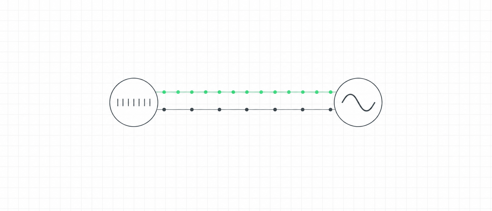
The heart and brain communicate in both directions. The brain adjusts heart rate through the autonomic nerves, and pressure sensors in the arteries report every beat back to the brainstem. The brain registers these signals, and the timing of a heartbeat can slightly change perception. The neurovisceral integration model links vagally mediated heart rate variability with prefrontal self-regulation, but the evidence is correlational and the links are small. The heart does not think.
Key points
- The brain sets the pace of the heart through the sympathetic and vagal nerves; the heart's own pacemaker supplies the basic rhythm.
- With each beat, pressure sensors in the large arteries signal the strength and timing of the heartbeat to the brain.
- The brain shows a measurable response to heartbeats, but the measurement is prone to artefacts and its meaning is still being worked out.
- Laboratory studies show that stimuli can be perceived differently depending on whether they arrive during or between heartbeats.
- The neurovisceral integration model is a model: it rests on correlations, and meta-analyses find small links between heart rate variability and self-regulation.
- The heart has a real network of nerve cells, but calling it a brain that thinks is an exaggeration.
- Physiological coherence is a measurable heart-rhythm pattern; claims that the heart's field influences other people are not established.
What is heart–brain interaction?
The heart and the brain are in constant two-way contact. The brain adjusts how fast and how strongly the heart beats; the heart, in turn, sends a stream of signals back with every beat. This page explains both directions, what the brain does with the heart's signals, and where the science stops and the popular claims begin. It separates three things that are often mixed together: established physiology, models that organise the evidence, and claims that are not supported.
How does it work?
Brain to heart. The heartbeat starts in the heart's own pacemaker, but its rate and force are regulated by the nervous system, hormones and other factors [S1]. The two branches of the autonomic nervous system do this work: sympathetic nerves speed the heart up, and the vagus nerve slows it down. Because the vagal brake acts quickly, it shapes the beat-to-beat changes measured as heart rate variability (HRV).
Heart to brain. The traffic also runs the other way. Each time the heart ejects blood, pressure sensors in the large arteries, the baroreceptors, signal the strength and timing of that beat to the brain; between beats they fall quiet [S2]. Together with other sensory nerve fibres from the heart, these signals give the brain a beat-by-beat report on the cardiovascular system. Breathing modulates this loop too, which is why heart rate rises and falls with each breath, the breath-linked heart rhythm.
How is it measured?
Responses to heartbeats. When brain activity is averaged around each heartbeat, a small response appears, called the heartbeat-evoked potential. Researchers use it to study how the cortex processes signals from the heart, and a review concluded that it can be a reliable measure only when artefacts from the heart's own electrical field and the pulse are carefully controlled [S3]. A meta-analysis found that the response grows with attention to the heartbeat and with arousal, differs in some clinical groups and is moderately linked to how well people sense their heartbeat, but it also warned that the reliability of these effects is unknown and that they may be driven by confounds [S4].
Timing within the heartbeat. A second approach presents a stimulus either during the heart's contraction (systole) or between beats (diastole). In one study, fearful faces were detected more easily and rated as more intense at systole, with stronger responses in the amygdala [S2]. In another, faint touches were noticed and located less often at systole [S5]. So the direction of the effect depends on the task: the heartbeat seems to sharpen some signals and dampen others. Both are single laboratory studies.
How these signals relate to body awareness in general is covered on the interoception page.
What affects it?
- Breathing. Breathing changes heart rate beat by beat and drives the largest swings in HRV, especially when slow; see how breathing changes HRV.
- Attention and arousal. Paying attention to the heartbeat and being aroused both change the brain's response to heartbeats [S4].
- Timing in the cardiac cycle. Whether a stimulus arrives during or between beats can change how it is perceived, in tasks studied so far [S2] [S5].
- Disease of the brain or heart. Acute brain injury can disturb the heart's control, as described below [S16].
What does the evidence show?
Established. The brain regulates heart rate and force through autonomic nerves and hormones [S1], and baroreceptors signal each heartbeat to the brain [S2]. The heart contains its own network of nerve cells [S11].
Context-dependent. The heartbeat-evoked potential is a usable research measure when artefacts are controlled [S3]. Physiological coherence, a smooth, regular heart-rhythm oscillation that appears when slow breathing brings breathing, blood pressure and heart rate into step, is a measurable pattern [S12]; the mechanics are on the breathing and HRV page, and how it differs from HRV itself is explained in HRV vs coherence.
A model, not a proof: neurovisceral integration. The neurovisceral integration model proposes that the same brain networks that help control attention and emotion, in particular inhibitory pathways from the prefrontal cortex through the amygdala to the brainstem, also adjust heart rate and therefore HRV [S6]. Its authors report that people with higher vagally mediated HRV tend to do better on executive-function tasks linked to the prefrontal cortex [S7]. Larger syntheses find a link, but a small one: across many studies, HRV and self-regulation showed only a weak association, with stronger results in published than unpublished studies, a sign of publication bias [S8]; a meta-analysis of correlational studies found a weak association between vagally mediated HRV and executive function [S9]; and a systematic review judged the number of studies too small to understand the relationship well [S10]. These are associations. They are consistent with the model, but they do not show that HRV causes better thinking, and they do not show that raising your HRV would improve it. A link this small says very little about any one person.
Emerging. Effects of the cardiac cycle on perception come from single laboratory studies with different tasks and opposite directions [S2] [S5]. The meaning of the heartbeat-evoked potential for body awareness is still uncertain [S4].
Unknown. Whether training heart rhythms changes brain function through these pathways has not been shown. Outcome evidence for training is on the HRV biofeedback page.
Does the heart have its own brain?
The heart does contain a real nervous system of its own: sensory, connecting and motor nerve cells that talk to nerve clusters in the chest and stay under the influence of the brain [S11]. Its main proposed job is local coordination of the heartbeat, and even that is framed by its leading researcher as a hypothesis [S11]; much of the detail comes from animal work. Calling it a "little brain" is a metaphor. Popular versions that say the heart has a mind of its own, that it thinks or that it holds intelligence are an exaggeration with no supporting evidence.
Does the heart's field affect other people?
Much of the popular material on heart coherence comes from the HeartMath Institute, whose researchers describe heart–brain pathways that can influence brain activity [S13] and hypothesise that magnetic fields from the heart help synchronise heart rhythms between people [S14]. HeartMath sells coherence-feedback devices, and the field idea is stated in its own papers as a hypothesis [S14]. Independent evidence that one person's heart field influences another person's body or mind is not established, and neither is the claim that "the heart thinks". The measurable part, a smooth heart-rhythm pattern during slow breathing, does not depend on either idea.
When heart–brain links matter in medicine
The connection is clearest in illness. Takotsubo (stress) cardiomyopathy is an acute heart failure syndrome that can follow an emotional or, more often, a physical trigger [S15]. After an ischaemic stroke, heart complications including arrhythmia are common in the first days, and disturbed autonomic control from the brain is the assumed mechanism [S16]. These are medical emergencies, not everyday stress effects.
What it does not tell you
- It does not make HRV a test of intelligence or self-control. The links are small and correlational [S8] [S9]; a single HRV reading says nothing about how well you think. Vagal tone cannot be measured directly; HRV measures such as RMSSD reflect vagally mediated changes in heart rate.
- It does not show that raising HRV improves the brain. That is a causal claim the neurovisceral evidence does not test [S7] [S10].
- A higher coherence score is not a brain reading. It describes the heart-rhythm pattern during breathing [S12], not prefrontal function or emotional state.
- Heartbeat effects on perception are small, task-specific and come from single studies [S2] [S5]; they are not a way to sharpen perception in daily life.
In ONDA
ONDA does not measure brain activity or the heart–brain connection. ONDA shows your pulse live during a practice, from the iPhone camera or an Apple Watch. A live coherence score, which needs the watch, is ONDA's own synchronization score for how smoothly your heart rhythm oscillates with your breath, a feedback metric rather than a clinical biomarker [S17]. ONDA does not diagnose anything. See what ONDA measures.
Educational information, not a diagnosis or medical treatment.
Evidence at a glance
| Claim | Evidence | Limitation |
|---|---|---|
| The heartbeat originates in the heart's own pacemaker, and heart rate and contractility are regulated by the nervous system, hormones and other factors. [S1] | Established | General physiology review; says nothing about any individual's heart. |
| At systole, arterial baroreceptors signal the strength and timing of each heartbeat to the brain; between beats they are quiescent. [S2] | Established | Background physiology stated in an experimental paper. |
| Neural responses to heartbeats, the heartbeat-evoked potential, are used to study how the cortex processes cardiac signals, and can be reliable only when artefacts are carefully controlled. [S3] | Context-dependent | Depends on artefact control; heterogeneous methods across studies. |
| A meta-analysis found moderate to large effects of attention, arousal and clinical status on the heartbeat-evoked potential and a moderate association with behavioural interoception measures, but the reliability of these effects is unknown and confounds may drive them. [S4] | Emerging | No standardised protocol; effects may reflect cardiac or somatosensory confounds. |
| In one study, fearful faces were detected more easily and rated as more intense at systole than at diastole, with larger amygdala responses. [S2] | Emerging | Single laboratory study in healthy volunteers; brief fear stimuli only. |
| In another study, touch stimuli were detected and localised less often during systole. [S5] | Emerging | Single laboratory study of threshold-level touch; a correction to the paper exists. |
| The neurovisceral integration model proposes inhibitory pathways from the prefrontal cortex to the amygdala and onward to the brainstem neurons that modulate heart rate and heart rate variability. [S6] | Debated | A model put forward by its authors; the pathways are inferred from anatomical and imaging work, not traced in each person. |
| The model links individual differences in heart rate variability with performance on executive-function tasks and prefrontal activity. [S7] | Debated | Mostly the authors' own studies; correlational. |
| Across many studies, greater heart rate variability was related to better top-down self-regulation, with a small effect. [S8] | Context-dependent | Correlational; stronger in published than unpublished studies, which suggests publication bias. |
| A meta-analysis of correlational studies found a small positive association between vagally mediated heart rate variability and executive functioning. [S9] | Context-dependent | Few correlational studies; moderated by the HRV measure and age. |
| A systematic review found that lower parasympathetic and higher sympathetic activity seem associated with worse cognitive performance, but the small number of studies limits conclusions. [S10] | Context-dependent | Observational studies in people without major disease; no causal inference. |
| The heart has an intrinsic nervous system of sensory, local-circuit and motor neurons that interacts with ganglia in the chest and is under the influence of central neuronal command. [S11] | Established | Much of the detailed physiology comes from animal research; its role in human thought or emotion is not shown. |
| The author of the 'little brain' description frames its coordinating role as a hypothesis. [S11] | Debated | Hypothesis in a review; concerns local cardiac control, not cognition. |
| Slow breathing near the resonant frequency brings breathing, blood pressure and heart-rate phases into step. [S12] | Context-dependent | Mechanistic review; the downstream benefits it names are hypotheses. |
| HeartMath-affiliated authors describe a coherence measure derived from heart rate variability and propose that the heart's magnetic field may mediate synchronisation between people, stated as a hypothesis. [S14] | Unknown | A hypothesis from an author affiliated with a company that sells coherence devices; not independently established. |
| HeartMath-affiliated authors describe heart–brain pathways through which afferent information can influence subcortical and frontal brain areas. [S13] | Debated | Narrative review with a commercial affiliation; 'can influence' is a possibility, not a measured effect size. |
| In a large registry, takotsubo (stress) cardiomyopathy followed emotional or physical triggers, physical ones being more common, and some patients had no evident trigger; it is an acute heart failure syndrome with substantial morbidity and mortality. [S15] | Established | Registry of hospital patients; not about everyday stress. |
| Cardiac complications including arrhythmia are frequent in the first days after an ischaemic stroke, and dysregulated central autonomic control is the assumed mechanism. [S16] | Established | The mechanism is assumed, not fully established; clinical setting. |
| ONDA's coherence score is derived from heart rhythm and breathing during a practice, needs an Apple Watch, and is a feedback metric rather than a clinical biomarker. [S17] | Context-dependent | Product documentation; describes the app, not any effect on the brain. |
Sources
- [S1] Gordan, Gwathmey & Xie (2015). Autonomic and endocrine control of cardiovascular function. World Journal of Cardiology. DOI 10.4330/wjc.v7.i4.204 · PMID 25914789
- [S2] Garfinkel et al. (2014). Fear from the heart: sensitivity to fear stimuli depends on individual heartbeats. The Journal of Neuroscience. DOI 10.1523/JNEUROSCI.3507-13.2014 · PMID 24806682 · Single laboratory study in healthy volunteers
- [S3] Park & Blanke (2019). Heartbeat-evoked cortical responses: underlying mechanisms, functional roles, and methodological considerations. NeuroImage. DOI 10.1016/j.neuroimage.2019.04.081 · PMID 31051293
- [S4] Coll et al. (2021). Systematic review and meta-analysis of the relationship between the heartbeat-evoked potential and interoception. Neuroscience & Biobehavioral Reviews. DOI 10.1016/j.neubiorev.2020.12.012 · PMID 33450331
- [S5] Al et al. (2020). Heart-brain interactions shape somatosensory perception and evoked potentials. Proceedings of the National Academy of Sciences of the USA. DOI 10.1073/pnas.1915629117 · PMID 32341167 · Single laboratory study; a correction was published (PNAS 2020, PMID 32690676); authors declare no competing interest
- [S6] Thayer & Lane (2009). Claude Bernard and the heart-brain connection: further elaboration of a model of neurovisceral integration. Neuroscience & Biobehavioral Reviews. DOI 10.1016/j.neubiorev.2008.08.004 · PMID 18771686 · Theoretical review by the authors of the model
- [S7] Thayer et al. (2009). Heart rate variability, prefrontal neural function, and cognitive performance: the neurovisceral integration perspective on self-regulation, adaptation, and health. Annals of Behavioral Medicine. DOI 10.1007/s12160-009-9101-z · PMID 19424767 · Narrative review mainly of the authors' own studies
- [S8] Holzman & Bridgett (2017). Heart rate variability indices as bio-markers of top-down self-regulatory mechanisms: a meta-analytic review. Neuroscience & Biobehavioral Reviews. DOI 10.1016/j.neubiorev.2016.12.032 · PMID 28057463
- [S9] Magnon et al. (2022). Does heart rate variability predict better executive functioning? A systematic review and meta-analysis. Cortex. DOI 10.1016/j.cortex.2022.07.008 · PMID 36030561 · Authors declare no competing interests
- [S10] Forte, Favieri & Casagrande (2019). Heart rate variability and cognitive function: a systematic review. Frontiers in Neuroscience. DOI 10.3389/fnins.2019.00710 · PMID 31354419
- [S11] Armour (2008). Potential clinical relevance of the 'little brain' on the mammalian heart. Experimental Physiology. DOI 10.1113/expphysiol.2007.041178 · PMID 17981929 · Review drawing largely on animal research
- [S12] Sevoz-Couche & Laborde (2022). Heart rate variability and slow-paced breathing: when coherence meets resonance. Neuroscience & Biobehavioral Reviews. DOI 10.1016/j.neubiorev.2022.104576 · PMID 35167847
- [S13] McCraty & Shaffer (2015). Heart rate variability: new perspectives on physiological mechanisms, assessment of self-regulatory capacity, and health risk. Global Advances in Health and Medicine. DOI 10.7453/gahmj.2014.073 · PMID 25694852 · Conflict of interest: the first author is affiliated with the Institute of HeartMath (PubMed affiliation), which sells coherence-feedback devices
- [S14] McCraty (2017). New frontiers in heart rate variability and social coherence research: techniques, technologies, and implications for improving group dynamics and outcomes. Frontiers in Public Health. DOI 10.3389/fpubh.2017.00267 · PMID 29075623 · Conflict of interest: the author is affiliated with the HeartMath Institute (PubMed affiliation), which sells coherence-feedback devices; the field claims are stated as hypotheses
- [S15] Templin et al. (2015). Clinical features and outcomes of takotsubo (stress) cardiomyopathy. The New England Journal of Medicine. DOI 10.1056/NEJMoa1406761 · PMID 26332547 · International registry compared with matched acute coronary syndrome patients
- [S16] Scheitz et al. (2018). Stroke-heart syndrome: clinical presentation and underlying mechanisms. The Lancet Neurology. DOI 10.1016/S1474-4422(18)30336-3 · PMID 30509695
- [S17] ONDA — product documentation: What ONDA measures. What ONDA measures and how it reads your signals.
Related
- ScienceInteroception: How the Brain Senses the Body
- ScienceThe Autonomic Nervous System: Sympathetic and Parasympathetic
- ScienceThe Vagus Nerve: What It Does, and What Is Myth
- ScienceHeart Rate Variability: What It Is, What It Reflects, What It Isn't
- ScienceHow Breathing Changes HRV — and Why Slow Breathing Raises It
- ScienceHRV Biofeedback: What the Evidence Shows
- GlossaryInteroception
- GlossaryVagus Nerve
- GlossaryHeart Rate Variability
- ArticleDoes HRV Predict How Fast You Think and React? What the Research Shows
- ArticleCoherent Breathing: The 6-Breaths-a-Minute HRV Sweet Spot
- ToolResonance Breathing Rate Finder
How ONDA Science pages are made: every number comes from one checked list of facts, every claim is mapped to its sources and graded by strength of evidence, and sources need a DOI or PMID (manufacturer documentation is used only for device facts).